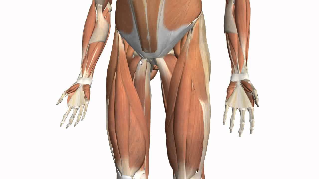
Upper Leg Tendon Anatomy : The upper leg muscles stock illustration. Illustration of muscles - 56286770. Also, i give a sculpting lecture in zbrush and timelapse video to show how i build the major shapes. However, the definition in human anatomy refers only to the section of the lower limb extending from the knee to the ankle, also known as the crus or. They're found on the ends of muscles, where they help. Quadriceps tendon to base of patella and onto tibial tuberosity via the patellar ligament action: You can read more about wrist tendons and the anatomy of the upper extremity, and view anatomy photos at www.handcare.org.
The lower leg is comprised of two bones, the tibia and the smaller fibula. Quadriceps tendon to base of patella and onto tibial tuberosity via the patellar ligament action: The thigh and leg bones articulate at the knee joint that is protected and enhanced by the patella bone that supports the quadriceps tendon. Tendon, tissue that attaches a muscle to other body parts, usually bones. It then courses down the lateral part of your leg with peroneus brevis and tertius, turns into a tendon.
Upper Back Muscle Chart - Building Back Muscles - 3 Mass Building Back Exercises ... - It is ... from i.ebayimg.com Tendons of the anterior compartment of the leg, the anterior tibial vessels, and the deep peroneal nerve pass under it. Tendon, tissue that attaches a muscle to other body parts, usually bones. There are several muscles which lie on the outside of your lower leg and are collectively known as the peroneal muscles (figure 1). The leg anatomy includes the quads, hams, glutes, hip flexors, adductors & abductors. To describe the mechanical properties of tendons. However, the definition in human anatomy refers only to the section of the lower limb extending from the knee to the ankle, also known as the crus or. There is no real division between the core and the upper leg; 1074 x 1856 jpeg 989 кб.
Fibula— a long, thin bone in the lower leg on the lateral side which runs along side the tibia from the knee to the ankle.
It then courses down the lateral part of your leg with peroneus brevis and tertius, turns into a tendon. Do anatomy tracings over those to find the leg bones. Information on the central tendon of the diaphragm by the anatomyzone daily feed. Tendon, tissue that attaches a muscle to other body parts, usually bones. It blends with the fibrous pericardium above, helping to. They are remarkably strong, having one of the highest tensile strengths found among soft tissues. The lower leg is comprised of two bones, the tibia and the smaller fibula. What is a peroneal tendon rupture? Medically reviewed by william morrison, m.d. It's the area that runs from the hip to the knee in each leg. Tendons are fibrous cords attached to muscles and bone. Iliotibial band syndrome description the iliotibial band is the tendon attachment of hip muscles into the upper leg (tibia) just below the knee to the outer side of the front of the leg. There are several muscles which lie on the outside of your lower leg and are collectively known as the peroneal muscles (figure 1).
See more ideas about leg anatomy, anatomy, anatomy drawing. Subscribe to learn interesting facts about the human body every day. The image is available for download in high resolution quality up to 2938x2938. Learn the origin/insertion, functions & exercises for the leg rotating your upper leg and pelvis to the inside or outside of your body's center line. Try to do both of these exercise on your own, before i post my answer briefly, it sits inside the quadriceps tendon and connects it to the front of the tibia by way of the patellar ligament.
Muscles of the Thigh Part 2 - Medial Compartment - Anatomy Tutorial - YouTube from i.ytimg.com Look for subcutaneous landmarks to figure out where the bones go. Try this movement out by standing on one foot with the other leg. Collectively, the muscles in this area plantarflex and invert the foot. Extends leg at knee in quad group. The thigh and leg bones articulate at the knee joint that is protected and enhanced by the patella bone that supports the quadriceps tendon. Lateral supracondylar line of femur, oblique popliteal ligament of knee insertion: The appendicular skeleton includes the bones of the shoulder girdle, the upper limbs, the pelvic girdle, and the lower limbs. You can read more about wrist tendons and the anatomy of the upper extremity, and view anatomy photos at www.handcare.org.
There are several muscles which lie on the outside of your lower leg and are collectively known as the peroneal muscles (figure 1).
Look for subcutaneous landmarks to figure out where the bones go. For more on tendon anatomy, refer here. Collectively, the muscles in this area plantarflex and invert the foot. This mri wrist coronal cross sectional anatomy tool is absolutely free to use. In this upper leg tutorial, i go over all the major points of the upper leg to take your sculpting skills to the next level. The human leg, in the general word sense, is the entire lower limb of the human body, including the foot, thigh and even the hip or gluteal region. Hands are outstretched, holding onto the handles of the bench. Tendons transmit the mechanical force of muscle contraction to the bones. The structure and composition of tendons allow for their linear region: Medically reviewed by william morrison, m.d. The thigh and leg bones articulate at the knee joint that is protected and enhanced by the patella bone that supports the quadriceps tendon. Learn the origin/insertion, functions & exercises for the leg rotating your upper leg and pelvis to the inside or outside of your body's center line. There are several muscles which lie on the outside of your lower leg and are collectively known as the peroneal muscles (figure 1).
There is no real division between the core and the upper leg; Tendons transmit the mechanical force of muscle contraction to the bones. Do anatomy tracings over those to find the leg bones. Subscribe to learn interesting facts about the human body every day. The thigh and leg bones articulate at the knee joint that is protected and enhanced by the patella bone that supports the quadriceps tendon.
Upper Limb Muscle Anatomy | 3D Anatomy with Actions of muscles, Forearm muscles, muscles of hand ... from i.ytimg.com The human leg, in the general word sense, is the entire lower limb of the human body, including the foot, thigh and even the hip or gluteal region. The anatomical basis of clinical practice. The lower leg is comprised of two bones, the tibia and the smaller fibula. Tendons are also bands of connective tissue. Tendons of the anterior compartment of the leg, the anterior tibial vessels, and the deep peroneal nerve pass under it. The pads of the machine are situated at the achilles tendon. Lateral supracondylar line of femur, oblique popliteal ligament of knee insertion: Want to learn more about it?
Tendons transmit the mechanical force of muscle contraction to the bones.
Tendons are fibrous cords attached to muscles and bone. The lower leg is comprised of two bones, the tibia and the smaller fibula. They're found on the ends of muscles, where they help. Quadriceps tendon to base of patella and onto tibial tuberosity via the patellar ligament action: Want to learn more about it? Medically reviewed by william morrison, m.d. Extends leg at knee in quad group. Learn vocabulary, terms and more with flashcards, games and other study tools. Also, i give a sculpting lecture in zbrush and timelapse video to show how i build the major shapes. See the pictures and anatomy description of knee joint bones, cartilage, ligaments, muscle and tendons with resources for knee problems & injuries. The pads of the machine are situated at the achilles tendon. However, many reflex pathways are also active in the legs and foot. 935 x 1601 jpeg 153 кб.
Share :
Post a Comment
for "Upper Leg Tendon Anatomy : The upper leg muscles stock illustration. Illustration of muscles - 56286770"


Post a Comment for "Upper Leg Tendon Anatomy : The upper leg muscles stock illustration. Illustration of muscles - 56286770"